Roxy came to LPVS as an emergency on Saturday 24th August – she had been out for a run and unfortunately collided her owners legs, which shockingly resulted in immediate paralysis of all her limbs. X-rays at her local vets raised concern for a broken neck and she was transferred to us by pet ambulance. Initial assessment by Colin Driver, RCVS specialist in Veterinary Neurology, suggested an injury to the spinal cord in her neck – but the nature of the trauma seemed low energy and so a fracture appeared unlikely. She was immediately anaesthetised for MRI and CT scans, which revealed a dislocation of the first two bones of the neck, squeezing the spinal cord – this had been caused by a congenital (from birth) malformation of the second bone which meant it did not articulate normally with its neighbour. This is a very uncommon finding in a dog of her breed – it is much more common in toy breed dogs such as Chihuahua’s and teacup Poodles. Considering the pressure on the spinal cord, emergency surgery was recommended. The dislocated bones were re-aligned from beneath, following a surgical approach through the throat. The joint was removed and bone graft was used to promote fusion. The joint was then stabilised with a total of 10 stainless steel threaded pins, which were enshrouded in a bolus of bone cement which sets solid after a few minutes. Post-operate CT scans confirmed the surgery was successful and Roxy was transferred back to wards to begin her rehabilitation. After several days, Roxy regained movement in all her limbs and before she went home was making attempts to stand and walk. We are due to re-examine her in a few weeks time to check on her progress. She was a lovely patient and we look forward to hearing about her progress.
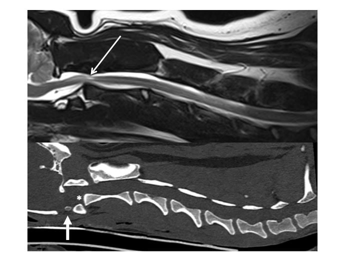
Figure 1
MRI and CT scans pre-operative show a severe spinal cord injury (open arrow) due to dislocation of the first two bones of the neck (atlantoaxial subluxation) – the second bone is malformed lacking a special process at the front called the dens (asterisk) that should connect to another piece of bone (closed arrow).
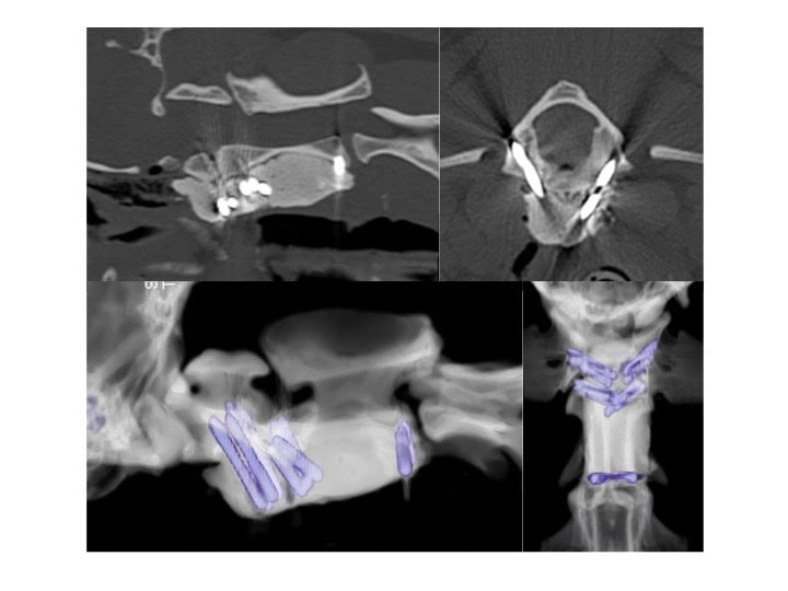
Figure 2
Post-operative CT scans demonstrating successful reduction of the bones and stabilisation with threaded pins and bone cement